Long case for Practical Examination
IP NO. 202109713
A 65 year old female from Alingapuram came to the OPD 25 days back with chief complaints of shortness of breath and swelling in both the legs
HOPI:
The patient was apparently asymptomatic 25 days ago when she developed:
SOB- she developed it while sitting and was sudden in onset, gradually progressive
( Grade IV )
Edema- started as b/l pedal edema and gradually progressed upto mid calf level
( Grade 2 ) associated with cough and fever.
Fever was low grade, continuous type. Relieved on medication.
No H/o chest pain , palpitations, decreased urine output , blood in urine, burning micturition.
Past History:
No history of similar complaints in the past.
Patient is a known case of Hypertension since 2 years for which she took medication irregularly.
Not a known case of Diabetes Mellitus, Asthma, TB, Epilepsy
H/o b/l knee pain since 1 year for which she was prescribed pain killers ( mostly NSAIDS )
No significant surgical history.
Family History: Not significant
Personal History:
Diet: mixed
Appetite : decreased
Sleep: reduced
Bowel and Bladder: regular
Addictions: none
Drug History: No known drug allergies.
General Examination:
The patient conscious, coherent, not co-operative.
Moderately built, moderately nourished.
Pallor- present
Edema- pitting type edema
(grade II ) involving both feet and legs upto mid calf.
Icterus- absent
Cyanosis : absent
Clubbing : absent
Koilonychia: absent
Lymphadenopathy: absent


Vitals:
Temperature: afebrile
Pulse Rate: 82 beats/min
Blood pressure: 130/ 80 mm hg
Respiratory Rate: 20 cycles/min
Spo2: 98%
Systemic examination:
Respiratory System:
Inspection of upper respiratory system-
- oral cavity- normal
- Nose- normal
- Pharynx- normal
Lower Respiratory Tract:
Inspection:
- trachea: central
- Symmetry of chest : symmetrical
- Movement: B/L symmetrical expansion of chest respiration
- No scars, engorged veins or sinuses.
Palpation:
All inspectory findings are confirmed by palpation.
- Trachea: central - confirmed by three finger test.

- Assessment of anterior and posterior chest expansion- B/L symmetrical expansion of chest.


- No chest wall tenderness
- Vocal fremitus- normal
Percussion : done in sitting position
Right | Left | |
Supraclavicular | Resonant | Resonant |
Infraclacicular | Resonant | Resonant |
Mammary | Resonant | Resonant |
Inframammary | Stony Dull | Stony Dull |
Axillary | Resonant | Resonant |
Infraaxillary | Stony dull | Stony dull |
Supra scapular | Resonant | Resonant |
Interscapular | Resonant | Resonant |
Infrascapular | Stony dull | Stony dull |
Auscultation:
Right | Left | |
Supraclavicular | NVBS | NVBS |
Infraclacicular | NVBS | NVBS |
Mammary | NVBS | NVBS |
Inframammary | Diminished | Diminished |
Axillary | NVBS | NVBS |
Infraaxillary | Diminished | Diminished |
Supra scapular | NVBS | NVBS |
Interscapular | NVBS | NVBS |
Infrascapular | Diminished | Diminished |
Vocal resonance: decreased over the basal areas
Basal crackles are present

Cardiovascular System :
Inspection :
- No scars sinuses and engorged veins.
- No visible pulsations
Palpation:
- apical impulse : heard in fifth inter coastal space
Auscultation:
- S1 and S2 heard
- No murmurs
Per Abdomen:
Inspection:
- Shape : elliptical
- Quadrants of abdomen moving in accordance with respiration.
- Umbilicus- central and inverted
- No scars sinuses or engorged veins
Palpation:
- No tenderness
- No organomegaly
Percussion
- tympanic
Auscultation:
- Normal bowel sounds heard
CNS:
- Higher mental functions-normal
- Cranial nerves- intact
- Sensory system- normal
- Motor system- normal
- Meningeal signs- absent
- Cerebellar signs- absent
Investigations:
1. Complete blood picture
- Hb 6.2 g/dl

2. Complete Urine examination
- albumin +2
3. Liver function Tests:

4. RBS: 82 mg/dl
5. Renal function Tests:
- Raised urea- 94 mg/dl
- Raised creatinine- 6.3 mg/dl
- Raised uric acid - 9.9 mg/dl

6. X-Ray

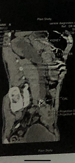
7. Ultrasound
- few subcentrimetric anechoic cysts in b/l kidneys
- Rt kidney- CMD lost. Grade III RPD
- Lt kidney- CMD partially maintained. Grade II RPD

8. ECG:

Interpretation of ECG:
• Rate- 100 beats/ min
• Normal sinus rhythm
• Normal axis
• p wave, qrs complex, t wave, pr interval, st segment - all appear to be normal
Provisional Diagnosis:
Kidney failure with bilateral plueral effusion
Treatment History:
- Inj piptaz 2.25 g iv TID

- TAB Lasix 40 mg PO/ BD

- TAB Nodosis 500 mg PO/BD

- TAB Orofer XT PO/OD

- TAB Shelcal CT PO/OD

- Inj Erythropoietin 4000 IU SC- twice weekly


- Salt and fluid Charting
- Vitals monitor and strict input/output charting.
Dialysis:
Patient underwent 6 sessions of dialysis since admission


Comments
Post a Comment