GM : PLEURAL EFFUSION
P.TRISHAALA


fatigue
breathlessness
lack of appetite and weight loss
vomiting
muscle cramping
swollen ankles and feet
puffiness around eyes
ROLL NO.121 8th SEMESTER
Case discussion about two cases of pleural effusion:
PLEURAL EFFUSION:
The accumulation of serous fluid within pleural space is termed pleural effusion.
In general , pleural fluid accumulates as a result of :
• Increased hydrostatic pressure/ decreased somatic pressure- TRANSUDATIVE EFFUSION. This is seen in cardiac, liver and renal failure.
• Increaed microvascular pressure due to disease of pleura or injury to adjacent lung- EXUDATIVE EFFUSION.
The common causes is pleural effusion are:
•Pneumonia ( para pneumonic effusion )
• Tuberculosis
• Pulmonary infarction
• Malignant disease
• Cardiac failure
• Sub diaphragmatic disorders ( subphrenic abscess, pancreatitis etc)
ref: Davidson's principles and practice of medicine
CASE 1:
A 54 year old male patient came to the OPD with chief complaints of
• Pain in the left side of chest, stabbing type,radiating to the left upper back since 2 days
• Difficulty in breathing since 2 days.
No h/o of palpitations, orthopnea, PND , vomittings
• History of burning sensation of both feet since 1 year.
* CHEST PAIN can be
• central
• peripheral
DD for Central chest pain:
• Cardiac
• Aortic
• Oesophageal
• Pulmonary embolus
• Mediastinal malignancy
•Anxiety/emotion
DD for Peripheral chest pain:
• Lungs/pleura
• Musculoskeletal
• Neurological
* BREATHLESSNESS may be due to
• Pulmonary edema
• Pulmonary embolus
• Asthma
• Exacerbation of COPD
• Pneumonia
• Metabolic acidosis
• Psychogenic
PAST HISTORY:
Pt lost sight in both eyes 10 years ago due to glaucoma.
Pt is not a known case of HTN , DM , asthma , TB, epilepsy, cardiovascular disease
EXAMINATION:
• Pt was conscious and coherent
• Virals
Temp - 100 F
Pulse- 104 bpm
BP- 160/100 mmhg
•CVS - S1 S2 heard. No murmurs
• P/A- soft, non tender
• RESPIRATORY SYSTEM:
Bilateral air entry +
Decreased Breath sounds in left ISA
Coarse crepitations heard
INVESTIGATIONS:
• Hemogram- showed increased TLC and neutrophil count- suggestive of infection
• Urine examination- increased sugars but negative for ketone bodies.
• RBS on presentation - 740 mg/dl
• RFT- normal expect slightly elevated phosphorus levels.
• Serum osmolarity increased
• ECG on day 1:

• CHEST X Ray on day 1- slight effusion on left

He was treated with Insulin and Glimeperide to control sugar levels.
•FEVER CHART:

•CHEST X Ray on day 4-

•HRCT:

Findings :
* left lower lobe consolidation
* right middle lobe peripheral consolidation
* moderate loculated left pleural effusion causing passive partial atelectasis of left lower lobe
* multiple small radio- opacities in gall bladder.
• Culture sensitivity of sputum showed positive foe
KLEBSIELLA PNEUMONIAE
• PLUERAL FLUID - negative for malignant cytology.
Smear showed- scanty lymphocytes and neutrophils against an esosinophilic background
Pleural sugar and LDH elevated
• CHEST X Ray day 6:
 |
• CHEST USG - thick septations and collapsed lung with minimal fluid.
PROBABLE DIAGNOSIS:
•Left loculated pleural effusion with consolidation of left lower lobe due to bacterial pneumonia ( klebsiella)
Or maybe viral in origin
• De novo Type 2 DM
• Cholelithiasis
TREATMENT:
• popped up position
• vital monitoring
• GBRS 4th hourly
• inj piptaz
• inj pan
• tab glimiperide
• inj neomol
• tab telma
• vit c
• Stricy I/O charting
• Surgical- breakage of loculations ans septations can be considered.
CASE 2
A patient came to the OPD with chief complaints of
• persistent cough
• shortness of breath (grade 3/4)
• lack of appetite
• left sided chest pain- non radiating and heaviness since 4 days
CASE HISTORY:
• bilateral edema upto knee since 2 years-pitting type, intermittent and relieved on medication.
• decreased urine output with burning micturition since 2 months.
It was associated with dribbling of urine, increased frequency, urgency and incomplete evacuation.
• bilateral joint pain in knee since 2 years
No complaints of palpitations, PND , orthopnea
No h/o fever
PAST HISTORY:
Not a known case of HTN, DM , Asthma , Epilepsy, TB, cardiovascular disease.
PERSONAL HISTORY:
• occasionally drinks alcohol
• was a beedi smocker- stopped 5 years ago
DRUG HISTORY:
History of NSAID Abuse
EXAMINATION:
Conscious coherent cooperative
Pallor +
Edema + b/l pedal edema extending upto the knee
( grade 3), pitting type.
VITALS:
Temp - Afebrile
Pulse- 84bpm
BP- 130/ 80 mmhg
CVS- S1 S2 hears. No murmurs
P/A- soft, non tender
CNS
• higher mental function and cranial nerves- intact
• sensory and motor system- normal
• reflexes - present
• no cerebellar or meningeal signs
RESPIRATORY SYSTEM
• vesicular breath sounds- decreases on left ISA
•no adventitious sounds
On patient’s previous visit to the hospital, the following investigations were done and the following treatment was given
INVESTIGATIONS:
•RBS 124 mg /dl
•Serum protein 6.2 g/DL
•Serum LDH 449 IU/L
•RFT
Urea : 61 mg /dl
Creatinine: 2.5 mg / dl
Uric acid- 8.4 mg/dl
Calcium 10.5 mg / dl
Phosphorus 3.1mg / dl
Sodium. 140 meq / lit
Potassium 4.2 meq/ lit
Chloride 103 meq/ lit
•Pleural fluid:
Protein : 4.2 gm / dl
Sugar :53 mg / dl
LDH 807 IU/L
•CUE :
Albumin :++
3-4 pus cells
2-3 epithelial cells
RBC nill
Sugars nil
•HEMOGRAM :
Hb: 11.6 g/dl
WBC:13,300 cells / cumm
RBC : 4.27 million/ cumm
platelets: 3.74lacks/cumm
Lymphocytes 16%
Smear : normocytic normochromic
ABG:
pH -7.45
Pco2 -23.8
Po2- 74.8
Hco3 -16.4
St.hco3- 19.7
O2sat -95.7
X-ray chest showed left sided pleural effusion
TREATMENT:
• tab lasix
• tab paracetamol
• inj neomol
• inj augmentin
• vital monitoring
• strict I/O charting
Therapeutic pleural tap was done- 500 ml fluid which was exudative in nature.
The patient came back to the hospital as his cough was persistent and sob was severe.
Presently the following investigations were done:
INVESTIGATIONS:
• Serum protein- 7.0 g/dl
•Serum LDH -263 IU / L
•Pleural fluid:
*Sugar- 74 gm/dl
*Protein- 6.2 gm/dl
*LDH- 476 IU/L
*VOLUME- 3 ml
*COLOUR -Pale yellow
*APPEARANCE- Hazy ( clot)
*TOTAL COUNT-1900 Cells/cumm
*DIFFERENTIAL COUNT
NEUTROPHILS- 02
LYMPHOCYTES -98
RBC-Nil
OTHERS -Nil
• X- Ray chest - showed neither progression nor regression of left sided pleural effusion.
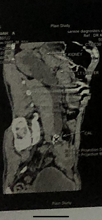
• CT CHEST- findings:
* centrilobular nodules with tree in bud appearance of
in apico posterior segment of left upper lobe, superior and basal segment of left lower lobe.
* loculated left pleural effusion with mild pleural thickening.
These findings are consistent with
• chronic infection, majorly tuberculosis
• secondaries of lung
Other findings:
*small right kidney- chronic kidney disease
*generalised increased bone density
PROBABLE DIAGNOSIS:
Chronic kidney disease stage 3b secondary to NSAID Abuse.
Left pleural effusion (exudative) secondary to:
• tuberculosis
• malignancy
* malignancy can be ruled out mostly as the radiologist repost say no mass found.
However it still needs to be evaluated and FNAC can be done.
* To confirm tuberculosis, sample was sent for CBNAAT and results are awaited.
PRESENT TREATMENT BEING GIVEN:
• inj optineuron
• tab pan
• tab lasix
• tab Ultracet
• fluid restriction
* if tuberculosis is confirmed, the patient should immediately put of anti tubercular therapy
* if the diagnosis is malignancy, the course of treatment depends on type and extent of disease.
NSAID ABUSE AND CHRONIC KIDNEY DISEASE :
Chronic kidney disease is characterised by gradual
loss of kidney function over time.
It includes conditions which damage the kidney and decrease its ability to work.
Over time wastes build up in the blood leading to various complications such as:
• high blood pressure
• anemia
• weak bones
• poor nutritional health
• nerve damage
The most causes of CKD are:
- Diabetes mellitus 20-40%
- Interstitial diseases- often drug induced 20-30%
- Glomerular diseases- IgA nephropathy 10-20%
- Hypertension 5-10%
- Systemic inflammatory diseases- SLE, vasculitis 5%
- Renovascular Disease 5%
- Congenital and inherited- Polycystic kidney disease and Alport's syndrome 5%
- Unknown 5-20%
Presenting complaints:
breathlessness
lack of appetite and weight loss
vomiting
muscle cramping
swollen ankles and feet
puffiness around eyes
dry and itchy skin
anemia
increased frequency of urination
raised urea and creatinine on blood examination

anemia
increased frequency of urination
raised urea and creatinine on blood examination
fall in GFR
Ref:Davidson's principles and practice of medicine

EFFECTS OF NSAID ABUSE ON KIDNEYS
Long term of use of Non steroidal anti inflammatory drugs is known to cause kidney damage:
Mechanism:
The kidneys receive 25% of cardiac output and are the major organs for drug excretion.
Due to this the renal arterioles and glomerular capillaries are extremely vulnerable to the affects of drugs.
NSAID are one of the most commonly used over the counter drugs.
NSAIDs inhibit the enzymes COX 1 and COX 2 which are rate limiting enzymes in the formation of prostaglandins and thromboxane.
“Adverse renal effects from these drugs are caused by two distinct pathological entities.
The first mechanism of acute kidney injury (AKI) from NSAIDs is due to reduced renal plasma flow caused by a decrease in prostaglandins, which regulate vasodilation at the glomerular level. NSAIDs disrupt the compensatory vasodilation response of renal prostaglandins to vasoconstrictor hormones released by the body. Inhibition of renal prostaglandins results in acute deterioration of renal function after ingestion of NSAIDs.
The second mechanism of AKI is acute interstitial nephritis (AIN), which is characterised by the presence of an inflammatory cell infiltrate in the interstitium of the kidneys”
Ref:
There are case studies as below which show comparatively early progression of CKD among patients who use NSAIDs for long durations.
2..
4.jeje5gm/IV/TI3.. Pan 40mg/IV/OD4. T. BD (2.5mg - .5mg)
5. T. Ultracet 1/2 tab QID
6. BP PR RR hourly
7. GRBS 4th hourly
8. T vitC 1000mg/OD
9. T Telma 40mg/od
10. Inj neomol 1gm/iv infusion if temp >101F
11. Strict I/O charting
1. Propped up position
2. Inj. piptaz 4.5gm/IV/TID day 7
3. Inj. Pan 40mg/IV/OD
4. T. Glimiperide BD (2.5mg - 1.5mg)
5. T. Ultracet 1/2 tab QID
6. BP PR RR hourly
7. GRBS 4th hourly
8. T vitC 1000mg/OD
9. T Telma 40mg/od
10. Inj neomol 1gm/iv infusion if temp >101F
11. Strict I/O charting


Comments
Post a Comment