GM : Case discussion: Pedal Edema
We were given 3 cases of pedal edema to analyse and understand the topic.


The complete cases can be found here:
CASE 1:
A 55yr old female patient came to the OPD with chief complaints:
• left sided pedal edema since 2 months
Pitting type
Progressive
• pain and swelling in left gluteal region since one month.
• pt is unable to perform daily activities due to pain
• Fever on and off since 1 month
•Underwent quack procedure 15 days ago for swelling.
• Developed pus points over gluteal area with discharge
after the procedure.
• pt consumes alcohol and cigarette since 10 years
• not a known case of HTN, DM, TB, Asthma, Epilepsy
• H/o of NSAID usage since 1 yr knee joint points
• H/o haemorrhoids and bleeding PR 2 months back.
UNILATERAL LEG SWELLING:
Most leg swelling is caused by oedema, the accumulation of fluid within the interstitial space.
The three explanatory mechanisms of edema are:
• increased hydrostatic pressure in the venous system due to increased intravascular volume or venous obstruction.
• decreased oncotic pressure secondary to a decrease in the plasma proteins that retain fluid within the circulation.
• obstruction to lymphatic drainage (lymphoedema)
All patients presenting with unilateral leg swelling should be evaluated for:
• Deep vein thrombosis
• gastrocnemius muscle tear
• ruptured baker’s cyst
• compartment syndrome
• cellulitis
• superficial thrombophlebitis
EXAMINATION:
Pt is conscious, coherent, cooperative
Pedal edema- left side , pitting type
Scars and pus points over left gluteal region
VITALS:
• temp- 99.8 F
• pulse -88bpm
•BP-110/80 mmhg
•RR-18 cycles/min
CVS ,RR ,P/A, CNS- normal on examination
INVESTIGATIONS:
• Hemogram:
*Hb- 8.8 g/dl
*Increased lymphocytes and nuetrophils- points towards infection
*Smear- microcytic hypochromic anemia with neutrophilic leukocytosis
•CUE - albumin +
•RFT- increased creatinine, urea, uric acid levels
•Venous Doppler- ruled out DVT
• LFT- increased ALP
decreased protein and albumin
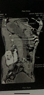
• USG abdomen
* abdominal distension
* grade 1 fatty liver
* grade 2 renal parenchymal injury
• serum creatinine- increased
• Reticulocyte count- increased
•Culture
* blood- negative
* urine- negative
* pus from gluteal abscess- klebsiella pneumoniae isolated.
PROBABLE DIAGNOSIS :
Acute kidney injury secondary to left lower limb cellulitis and left gluteal abscess.
TREATMENT:
• Inj metrogyl
• inj vancomycin
•tab pantop
• lower limb elevation
• regular dressing
ACUTE KIDNEY INJURY:
It is sudden, often reversible loss of renal function, which develops over days or weeks and is often accompanied by reduction in urine volume.
Pathophysiology
• pre-renal: perfusion you kidney is reduced
• renal: primary insult affects kidney itself
• post renal: obstruction to urinary flow
AKI SECONDARY TO LOWER LIMB CELLULITIS:
AKI occurs commonly in hospitalized patients with multiple comorbidities.
Such patients lead to a broad differential diagnosis for AKI including prerenal AKI, acute tubular injury/acute tubular necrosis, infection-related GN, and drug-induced acute interstitial nephritis.
Epidemiologically, the two most likely causes of AKI in the hospitalized patient are prerenal AKI and acute tubular injury/necrosis (ATI/ATN).
Prerenal or hemodynamic AKI may result from either true or effective volume depletion, especially in patients such as this who have underlying CKD and are receiving renin-angiotensin system blockers.
Ischemic and nephrotoxic ATI/ATN are common causes of AKI.
Acute PIGN from MRSA cellulitis and bacteremia should be strongly considered as a potential cause of AKI. MRSA infection is well described to promote an immune-complex GN in hospitalised adults
CASE 2:
An 18 yr old male patient came to the OPD with chief
complaints of
• tingling and numbness of both legs since 15 days
• tingling and numbness of both hands since 4 days
HOPI
patient was apparently asymptotic 15 days ago when:
• sudden onset of loss of sensation in both legs
• sudden onset of bilateral pedal edema - subsided with medication
• difficulty in wearing chappal since 10 days
• difficulty in climbing stairs since 8 days
• difficulty in squatting since 6 days
• later upper limb involvement was present with electric shock like sensations- both distal and proximal
•at time of presentation unable to stand or walk without support
• difficulty in eating, buttoning of shirt and combing of hair.
No h/o breathing difficulty
Limbs are flaccid and loose
No wasting/thinning of muscle
No h/o pain, fatigue, muscle cramps , fasciculations, twitching of muscles, involuntary movements.
Able to feel cloth
Able to differentiate hot and cold
No h/o fever ,vomiting , diarrhoea, sore throat.
Based on history, provisional diagnosis of Guillain Barre syndrome was made.
EXAMINATION:
Pt is conscious, coherent, co operative.
Temp- afebrile
Pulse- 54 bpm
BP- 100/70 mmhg
CVS, RS, P/A - normal on examination
CNS
• higher mental functions and cranial nerves-intact
• Sensory system - normal
• no cerebellar and meningeal signs
• motor:
*bulk- Normal
* tone: decreased in bilateral upper and lower limbs
* power: reduced in muscles forearm and wrist joint
* superficial reflexes - present
* deep tendon reflexes- absent
Based on history, provisional diagnosis of
Guillain Barre syndrome with acute motor and sensory axonal neuropathy was made
INVESTIGATIONS:
• Hemogram- Normal
• RFT- Increased creatinine, calcium , phosphorus
• LFT- increased total and direct bilirubin
•Viral serology - negative
• Nerve conduction study was suggested- indicated severe axonal injury.
• Nerve biopsy can be done
ADVISE ON DISCHARGE:
• Physiotherapy to all 4 limbs
• B complex tablet
GUILLAIN BARRE SYNDROME:
Guillain-Barré syndrome (GBS) is a rare neurological disorder in which the body's immune system mistakenly attacks part of its peripheral nervous system—the network of nerves located outside of the brain and spinal cord. GBS can range from a very mild case with brief weakness to nearly devastating paralysis, leaving the person unable to breathe independently.
The exact cause of GBS is not known. Researchers don’t know why it strikes some people and not others. It is not contagious or inherited.
SYMPTOMS:
Unexplained sensations often occur first, such as tingling in the feet or hands, or even pain (especially in children), often starting in the legs or back. Children will also show symptoms with difficulty walking and may refuse to walk. These sensations tend to disappear before the major, longer-term symptoms appear. Weakness on both sides of the body is the major symptom that prompts most people to seek medical attention. The weakness may first appear as difficulty climbing stairs or with walking. Symptoms often affect the arms, breathing muscles, and even the face, reflecting more widespread nerve damage. Occasionally symptoms start in the upper body and move down to the legs and feet.
DIAGNOSIS:
Key diagnostic findings include:
- Recent onset, within days to at most four weeks of symmetric weakness, usually starting in the legs
- Abnormal sensations such as pain, numbness, and tingling in the feet that accompany or even occur before weakness
- Absent or diminished deep tendon reflexes in weak limbs
- Elevated cerebrospinal fluid protein without elevated cell count.This may take up to 10 days from onset of symptoms to develop.
- Abnormal nerve conduction velocity findings, such as slow signal conduction
- Sometimes, a recent viral infection or diarrhea.
TREATMENT:
There is no known cure for Guillain-Barré syndrome. However, some therapies can lessen the severity of the illness and shorten recovery time.
CASE 3:
A 65 year old female patient came to the OPD with complaints of
• abdominal distension since 4 days
• diffuse abdominal pain since 4 days- spasmodic ,
non radianting
• decreased urination since 4 days
• burning micturition since 4 days
• constipation since 2 days
• grade 2 dyspnea
• h/o fluid loss 4 days ago ( 2 episodes of vomiting )
• dyspnea grade 2
• bilateral pedal edema upto knee
Patient is a known case of
• Diabetes since 6 months- is on metformin
• HTN since 3 years- is on anti hypertensive therapy
( tab atenolol and tab amplodipin)
EXAMINATION:
Patient is conscious coherent and cooperative
Vitals
•Temp- afebrile
• Pulse- 84 bpm
• BP- 110/70 mmhg
•RR- 16 cycles/ min
CVS , CNS - normal on examination
RESPIRATORY SYSTEM:
• bilateral air entry +
• trachea- central
• wheeze- absent
• dyspnea- grade 2
• breath sounds- vesicular
• adventitious sounds - b/l coarse crepitations
P/A
• soft , no tenderness
• no palpable mass
• no free fluid , no bruits
• liver spleen- not palpable
• bowel sounds- sluggish
INVESTIGATIONS:
• Hemogram- Normal
• RFT - increased urea, creatinine, uric acid
• LFT- * increased total and direct bilirubin
* increased liver enzymes
* decreased albumin and protein
• Bleeding time and clotting time are on the higher end
• Urine culture- positive for E.Coli
• CHEST X Ray- left lung Lower lobe collapse
PROBABLE DIAGNOSIS
•Acute kidney injury secondary to Urinary tract infection with hypoalbuminemia
•Urinary tract infection
• Left lung lower lobe collapse
• Grade 2 fatty liver
• Type 2 DM
The patient underwent dialysis indication being anuria
and increased blood urea and serum creatinine levels
ADVICE:
• salt restriction
• tab nodosis
• tab aldatone
• tab amlong
• tab shelcal
• syrup potchlor
* hypoalbuminemia which was secondary to fatty liver
caused decreased plasma oncotic pressure leading to fluid accumulation ( ascites and pedal edema )
* UTI lead to an ascending infection leading to pyelonephritis and acute kidney failure.
* Kidney failure leads to increased levels of toxins to build up which affected the lungs leading to left lower lobe collapse
The kidneys receive more cardiac output on a per-gram basis than some other organs such as the liver (approximately 25% of cardiac output). Therefore, kidneys are constantly exposed to small peptides and immune regulatory molecules, which can reabsorb these substances from circulation and excrete them.
The harmful effects of AKI on the lung function could relate to the loss of normal balance of immune, inflammatory and soluble mediator metabolism.The kidney plays a key role in cytokines metabolism and clearance. Impaired kidney function is associated with cytokine imbalance (both production and elimination) in the circulation. It revealed that an important pathway of lung injury subsequent kidney injury could arise from cytokine dysregulation in the kidney, with further activation of the lung's indigenous immune cells and respiratory complications

Comments
Post a Comment