General medicine case presentation: HEMIPARESIS
P.TRISHAALA REDDY


ROLL NO.121 8th semester
This week we were given 3 cases to analyse and understand the topic of hemiparesis.
The complete cases can be found here:
HEMIPARESIS or unilateral paresis is is the weakness of one entire side of the body.
Common causes are:
• Vascular: cerebral hemorrhage, stroke
•Infective: meningitis, encephalitis, brain abscess, spinal epidural abscess
•Neoplastic: glioma, meningioma, brain and spinal cord tumors
• Demyelination: multiple sclerosis, ADEM, nueromyelitis optica
•Traumatic: epidural subdural hematoma, vertebral compression fracture.
•Iatrogenic: local anaesthetics rapidly given Inta-arterially.
•Ictal: seizures, Todd’s paralysis
• Congenital: cerebral palsy, neonatal onset multisystem inflammatory disease (NOMID)
•Degenerative: ALS, corticobasal degeneration.
•Parasomnia: sleep paralysis
CASE 1:
A 45 year old female patient came to the OPD with the following complaints:
• headache since the past 2 years
•b/l lower limb weakness since 5 months
•h/o body pains, disturbed sleep, auditory hallucinations, double vision.
Diagnosed as depression without psychotic features.
Put on bulotin, ehisan’s plus for 1 week
She had sudden fall- slurring of speech and and swaying present.
Unable to get up from bed. Giddiness On lying down
CT was done- showed small calcific foci in left high parietal region- Diagnosed as Ischemic stroke.
PAST HISTORY:
• patient is a known case of hypothyroidism and is on Tab. Thyronorm since the past 4 months
•h/o uterine prolapse 20 yrs ago for which hysterectomy was done.
• her 2nd child died at 1 month of age due to unknown reasons
• her 3rd child died at age 21 due to kidney failure.
( Reason for her depression?? )
DRUG HISTORY:
Nortryptiline
Cerebroprotein
MVT
Ecosprin
Clopitab
Atovastatin
NSAIDS
EXAMINATION:
Patient conscious, coherent, cooperative.
On CNS EXAMINATION:
•Higher mental function- normal
•Oriented to time place and person
•Cranial nerves- 3rd and 6th CN affected
7th cranial nerve on affected on left side
•Motor system:
Hypotonia in right upper and lower limb
Reflexes-Superficial reflex:
Abdominal reflex absent
Plantar Extensor reflex present
Deep reflex: exaggerated
• Gait- ataxic
• Sensory system- Normal
• No meningeal signs
INVESTIGATIONS:
•Hb- 11.4 gm/dl
•ECG
•MRI plains and contrast with angiogram- patchy hyperintensities on both cerebral hemispheres.
MR angi: hypoplastic basilar-vertebral arteries
•Carotid Doppler - soft plaque in left carotid bifurcation causing no significant stenosis
TREATMENT:
• inj methylprednisolone
• Inj Zofer
• tab PCM
• tab atorvastatin
• physiotherapy
• BP charting 4 hrly
DIFFERENTIAL DIAGNOSIS:
• Neuro Behçet’s disease
• Multiple sclerosis
• Neuromyelitis optica
• Vasculitis
• Nuerosarcoidosis
*NEURO BEHÇET’S DISEASE
“Behcet's disease (BD) is an autoimmune disease characterized by recurrent oral and genital ulcers and uveitis. The exact pathogenesis is still unclear, but the main histopathological feature is of widespread vasculitis of arteries or venules of any size.
Neurological involvement may occur in the course of the disease and have two major forms: vascular and parenchymal . Parenchymal lesions lead to inflammatory lesions in the brain stem, diencephalon, basal ganglia and less frequently, the spinal cord and cerebellum.
cellularity are found in 60% of patients .The second form of neurological involvement is dural sinus thrombosis mainly characterized by headache and papilloedema. ”
*NEURO MYELITIS OPTICA:
“Neuromyelitis optica (NMO) is an autoimmune inflammatory central nervous system disorder typically associated with longitudinally extensive transverse myelitis (LETM) and optic neuritis. The serum autoantibody against aquaporin-4 (anti-AQP4 antibody) is a highly specific disease marker and is the basis for establishing the concept of NMO spectrum disorder (NMOSD). Isolated involvement of the spinal cord, optic nerve, brainstem, or even cerebrum can be seen in NMOSD”
Given in the link below are two cases of NMO presenting as isolated white matter lesion complaining of unilateral weakness.
* MULTIPLE SCLEROSIS:
"Multiple sclerosis (MS) affects the brain and spinal cord. Early MS symptoms include weakness, tingling, numbness, and blurred vision. Other signs are muscle stiffness, thinking problems, and urinary problems. Treatment can relieve MS symptoms and delay disease progression. It is a long-lasting disease that can affect your brain, spinal cord, and the optic nerves in your eyes. It can cause problems with vision, balance, muscle control, and other basic body functions."
Multiple Sclerosis (MS) Center: Symptoms, Treatments ...www.webmd.com › multiple-sclerosis
Here is an article about cases of MS presenting with hemiparesis
Hemiparetic multiple sclerosis - Journal of Neurologyjnnp.bmj.com › content › jnnp › 675.full.pdf
* NUEROSARCOIDOSIS
"Neurosarcoidosis is an uncommon but potentially serious manifestation of sarcoidosis. While the cranial nerves are most frequently affected, neurosarcoidosis can involve other nervous system tissues including the meninges, brain parenchyma (especially the hypothalamic region), spinal cord, peripheral nerve, and muscle. Diagnosis may be particularly challenging when neurosarcoidosis occurs in isolation. Diagnostic criteria usually include histologic identification of a noncaseating granuloma, supportive laboratory or imaging tests or both, and a compatible clinical course. Treatment has not been subjected to rigorous study, but corticosteroids are typically the first line of therapy and approximately half of patients have substantial benefit. For patients who are refractory to or intolerant of corticosteroid therapy, second-line agents include azathioprine, methotrexate, cyclosporine, cyclophosphamide, mycophenolate, and even cranial irradiation. The combination of infliximab and mycophenolate mofetil is under study as well."
CASE 2 :
A 35yr old female patient came to the OPD with a chief complaint of
• Vomitings ( 3 episodes ) 6 days ago followed by altered sensorium.
No h/o Truma, fever with cough and cold , chest pain
PAST HISTORY:
Known case of Tuberculosis 4 yrs back for which she took anti tubercular treatment for 6 months.
No other significant past or family history.
EXAMINATION:
Pallor- present
Mild dehydration and malnutrition present.
Doll’s eye - positive
Pulse- 110 bpm
RR- 20 cycles/min
BP- 100/60 mmhg
RBS- 96 %
CVS , Respiratory and GI systems are normal on examination.
CVS EXAMINATION:
• Higher mental function- pt is unconscious and stuporous.
• Cranial Nerves:
* 1st and 2ns - not elicited
* 3rd 4th 6th - normal
* 5th-Motor - not elicited
Sensory- not elicited
Corneal and conjunctival reflex- normal
* 7th - nasolabial fold- N and no deviation of mouth.
Corneal and conjunctival reflex- normal
Secretomotor- moistness of tongue and eyes- N
Sensory- not elicited
*8th - Renne’s ans Weber’s- not elicited
*9th and 10th- uvula centrally placed
Gag reflex present
*11th- trapezius and sternocliedomastoid - not elicited
* 12th- tongue tone normal
No wasting, no deviation.
• Motor system:
Decreased tone in right upper and lower limbs.
Power -not elicited in rt UL and LL. 3/4 in left UU and LL
Reflexs present
Primitive reflex- absent
Involuntary movements- absent
* Left lower limb shows continuous and intermittent movements.
• Sensory system- not elicited.
Pain in all four limbs
•Meningeal signs- neck stiffness present
•Cerebellar signs- absent
INVESTIGATIONS:
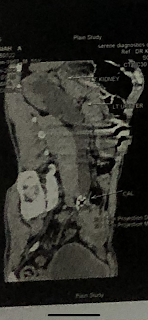
•MRI : CAPSULOGANGLIONIC HEMORRHAGE.
 |
| Add caption |
ANTIHCV ANTIBODIES: nonreactive
HIV : non reactive
HEMOGRAM :
28/4 29/4. 1/5. 3/5
HB 5.4 6.3. 7.1. 9.4
Platelets 1.94. 0.31. 1.11. 1.5
TLC 12000
PT. 20. 16
INR. 1.7. 1.1
APTT. 34
BT. 10. 2
CT. 15. 4
TO.BIL. 1.03. 1.48
DI.BIL. 0.27. 0.33
SGOT. 17. 20
SGPT. 8.8. 12
ALK.PH. 68. 64
TO.PRO. 7. 7.1
ALB. 4.1
A/G RATIO. 1.37
RFT
UREA. 17. 52
CREATININE. 0.89. 0.8
URIC ACID 2.6
CALCIUM. 10.5
PHOS. 4.2
SODIUM. 146
K+. 3.6
CL-. 100
COURSE IN HOSPITAL:
After admission to ICU:
•high grade fever with leukocytosis - managed with IV antibiotics.
• mass lesion ans hematoma
• recovered from coma spontaneously and mobilised herself slowly with persistent neurological deficits.
• still no speech due to affection of Broca’s area.
Psychiatry referral suggested in view of cognitive and higher mental functions.
“According to informent :-
C/o not talking only responding at times.
Stressors(financial, family, personal)
Crying spells + low mood+ she was active at work
GAB patient is lying on bed unable to move her right arm and leg
ETEC +, built not sustained
PMA decreased
Rapport could not be established
SPEECH:not uttering words but responding to sounds at times non verbal communication +at times though a bit slow
AFFECT: dysphoric
Further MSE could not be elicited , Orientation could not be assessed
Patient power gradually improved from 0/5 on right side to 3/5 and on left side improved for 3/5 to 4/5.
DIAGNOSIS:
• Cardiovascular accident causing right sided Hemiplegia-
Acute Hemorrhage involving left corona radiata and Lentiform nucleus, internal capsule with intraventricular extension.
Secondary to 1) AV malformations??
2) Underlying mass lesion with old pulmonary koch’s
TREATMENT:
• inj amikacin
• inj ceftrioxone
• tab pan top
• tab Zofer
• tab paracetamol
• b complex
• vitals monitoring
• DVT stocking, air bed and constant change of position
• tab fluoxetin
• chest , upper limb, lower limb physiotherapy
CVA- ACUTE HEMORRHAGIC STROKE causing HEMIPLAGIA:
A stroke occurs when the flow of blood to part of the brain is cut off or significantly reduced. Without the oxygen carried by the blood, brain cells can die quickly, which can cause permanent brain damage.
There are two kind of strokes: Hemorrhagic and Ischemic.
An ischemic stroke is caused by lack of blood flow to brain tissue. This can happen when the arteries in the brain narrow due to a condition such as atherosclerosis or thrombosis. Another cause of ischemic strokes is an embolism.
A hemorrhagic stroke is also called an intracerebral hemorrhage. An ICH occurs when a blood vessel ruptures and blood accumulates in the tissue around the rupture.
The two common causes of rupture are
• Aneurysm
• AV malformations
Presenting symptoms of hemorrhagic stroke:
• total or partial loss of consciousness
• nausea and vomiting
• numbness of one side of body
• seizures
• loss of balance
• dizziness
• problems in speech and swallowing
• disorientation and confusion
Usual treatment is to reduce blood pressure and slow down bleeding.
It can be managed by
• IV fluids
• Rest
• Management of other medical illnesses
• Speech, physical and occupational therapy.
• In severe cases surgery is done to repair the affected blood vessels.
CASE 3
A 65 yr old female patient came to the OPD with a complaints of
• loose stools since 2 days-10ep/day. Non-mucoid,
non-blood stained, not assc with pain abd.
• Fever since 2 days - low grade, intermittent, not assc with chills and rigour.
H/o one episode of vomitting- contained of food particles.
• Decreased urine output since 2 days. No burning micturition.
• Facial puffiness since 1 day
PAST HISTORY:
K/c/o
• right upper and lower limb weakness and slurring of speech since oct 2019 secondary to an infarct in the right parietal regions for which she is on anti platelet medication.
• HTN since 8 months- for which she is using medication.
No other significant past and family history.
EXAMINATION:
Vitals
• Pulse - 67 bpm
• RR- 20 cycles/min
• GRBS- 147 mg%
• spO2 - 96%
• BP- 50/20 mmhg at the time of presentation.
After resuscitation with IV fluid increased to 80/50 mmhg.
Respiratory, CVS and GI systems showed no abnormalities on examination
CNS Examination:
Higher mental function - normal
Cranial nerves-intact
Sensory system- normal
No cerebellare and meningeal signs
Motor system- power decreased (4/5) in all four limbs.
Reflexes- present
INVESTIGATIONS:
•CBP- hb- 4.5 gm% -
RBC - 1.6 mill/cumm
DLC- Polymorphs- 80% ( increased)
•PERIPHERAL SMEAR- microcytic hypochromic anaemia
•RFT- increased levels of urea creatinine and uric acid
•LFT-increased levels of SGOT and alkaline phosphatase.
Decreased protein and albumin.
•ABG
•USG- no sonological abnormality detected
•RBS- 99 mg/dl
•ECG
DIAGNOSIS-
AKI secondary to GASTROENTERITIS with METABOLIC ACIDOSIS.
HYPOVOLEMIC SHOCK with ANEMIA
Pre existing HTN and CARDIOVASCULAR ACCIDENT. ( Rt UL and LL weakness )
TREATMENT:
• IV fluids
• oral fluids
• oral rehydration therapy after each loose stool
• inj PAN
• inj zofer
• tab ecospirin
• vital monitoring
• inj lasix
• blood transfusion
ACUTE KIDNEY INJURY SECONDARY TO GASTROENTERITIS:
“Acute kidney injury (AKI), previously known as acute renal failure (ARF), characterized by sudden impairment of kidney function resulting in retention of nitrogenous and other waste products normally cleared by kidneys. AKI is not a single disease but, rather a heterogenous group of condition that share a common diagnostic features, especially increase in the blood urea nitrogen concentration and/or increase in plasma or serum creatinine concentration, often associated with reduction in urine volume.
AKI following volume depletion due to gastrointestinal fluid loss is common in India. Diarrheal diseases are common in India due to poor socioeconomic conditions, poor access to treatment, ignorance about personal hygiene, overcrowding, and climatic conditions which supports the propagation of infection”

Comments
Post a Comment