GENERAL MEDICINE CASE PRESENTATION: Patients presenting with shortness of breath
PATIENTS PRESENTING WITH SHORTNESS OF BREATH
P. TRISHAALA REDDY
ROLL NO.121 8TH SEMESTER
This week we were given 3 cases of patients presenting to the OPD with shortness of breath
https://medicinedepartment.blogspot.com/2020/05/re-case-based-online-learning.html?m=1
SOB or medically known as dyspnea is often described as intense tightening in the chest, air hunger, difficulty in breathing or a feeling of suffocation.
DIFFERENTIAL DIAGNOSIS FOR A PATIENT PRESENTING WITH DYSPNOEA:
Patients with breathlessness present either with chronic exertional symptoms or as an emergency with acute breathlessness, when symptoms can be prominent even at rest.
CAUSES OF DYSPNOEA:
- CARDIOVASCULAR
Acute dyspnoea : Pulmonary edema
Chronic exertional dyspnoea : Heart failure
Myocardial ischemia
- RESPIRATORY
Acute dyspnoea: Acute severe asthma
Acute exacerbation of COPD
Pneumothorax
Pulmonary embolus
ARDS
Inhaled foreign body
Lobar collapse
Laryngeal edema
Chronic exertional dyspnoea : COPD
Chronic Asthma
Lung Cancer
Interstitial lung disease
Chronic Pulmonary thromboembolism
Lymphangitis carcinoma
Large plural effusion
- OTHERS
Acute dyspnoea : Metabolic acidosis
Psychogenic
Hyperventilation
Chronic exertional dyspnoea : Severe anemia
Obesity
Deconditioning
Ref: Davidson's Principles and Practice of Medicine
a-case-of-dyspnoea-10-638.jpg
CASE 1:
https://madhur116.blogspot.com/2020/05/on-1452020.html?m=1
A 35 yr old male patient presented in the OPD with chief complaints of:
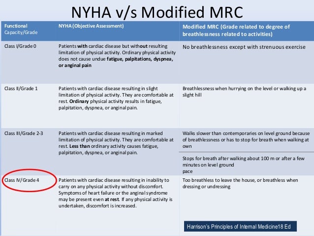
Shortness of breath since 2 weeks ( NYHA 3)
Pedal edema since 2 weeks - upto knee, pitting type
Paroxysmal nocturnal dyspnoea and weakness since 2 weeks
Patient gave a history of fever associated with chills 1 month back for which he was treated with antimalarials and symptomatically.
No significant past history
Drinks alcohol occasionally
BY HISTORY
- We can say that dyspnoea is more of chronic exertional type
- We can rule out respiratory causes as characteristic symptoms such a cough, wheezing, chest pain are absent.
- The symptoms are more consistent with a cardiac cause as SOB with pedal edema and PND are usually seen when there is a cardiac pathology.
EXAMINATION:
Pallor ,icterus, cyanosis , clubbing, lymphadenopathy absent
Edema present upto knee ( grade 2 )
Vitals : temp- Afebrile
BP- 130/80 mmhg
PR: 80 bpm
CVS - S1 S2 heard
RS- right ISA inspiratory crepts +
P/A- soft non tender
CNS- normal
JVP- raised
ON INVESTIGATION:
CBP- within normal limits
FBS- 102 mg/dl
PLBS: 205 mg/dl
Since a cardiac cause is suspected we do a 2D echo:
Findings: EF-27% ( REDUCED ejection fraction)
IVC Dilated
Mild TR +
Severe MR+
Trivial AR +
All chambers are dilated
Global hypokinesia
Severe LV dysfunction
Mild PAHT
No MS/AS
No PE/LV clot
All investigations are consistent with HEART FAILURE with reduced ejection fraction due to DILATED CARDIOMYOPATHY
CAUSES OF DILATED CARDIOMYOPATHY:
Idiopathic- 50%
Myocarditis- 9%
Ischaemic heart disease -7%
Infiltrative disease- 5%
Hypertension- 4%
HIV infection- 4%
Connective tissue disorder- 3%
Doxurubicin- 1%
Others- 10%
DIAGNOSIS: This may be a case of heart failure with dilated cardiomyopathy secondary to viral myocarditis with de novo DM type 2
Common virus involved in causing myocarditis are ADENOVIRUS, ENTEROVIRUS, COXSACKIEVIRUS, CMV, EBV etc
The clinical spectrum of viral cardiomyopathy can be classified as fulminant, acute, or chronic. Viremia is followed by cardiomyocyte infection. In the first phase, acute infection of cardiac myocytes results in myocyte death and activation of the innate immune response, including interferon gamma, natural killer cells, and nitric oxide. Antigen-presenting cells phagocytize released viral particles and cardiac proteins and migrate out of the heart to regional lymph nodes.
Under neurohumoral stimulation and hemodynamic stress, the ventricles dilate, leading to chronic cardiomyopathy.
Diagnosis and Treatment of Viral Myocarditis - NCBIwww.ncbi.nlm.nih.gov › pmc › articles › PMC2770911

1-s2.0-S1875213609001685-gr1.jpg
Further investigations that can be done:
ECG
Serum levels of troponin to rule out any ischaemic cause
Creatinine Phosphokinase fractions
Endomyocardial biopsy
Viral serology
PCR for detection of viral genome
TREATMENT:
The patient is already on:
Tab lasix
Tab isosorbide mononitrate
Tab hydralazine
Tab Telma
Tab Metformin
Fluid restriction
Salt restriction
Additional medications that can be added:
ACEi (enalapril)
Newer modalities of treatment for heart failure:
ARNI- combination drug of Angiotensin receptor blocker ( valsartan) and neprilysin inhibitors (Sacubitril) have known to show better efficacy than ACEi
Efficacy of Angiotensin Converting Enzyme Inhibitors and ...www.ncbi.nlm.nih.gov › pmc › articles › PMC6426571
CASE 2:
https://himabindu5.blogspot.com/2020/05/hello-everyone.html
A 55 yr old female patient came to the OPD with chief complaints of
Palpitations since 2 months
Chest pain since 2 months
Associated with shortness of breath (NYHA grade 3/4) and Paroxysmal nocturnal dyspnoea
Bilateral pedal edema upto ankle since 1 week
Decreased urine output since one week
no history of fluid loss
no history of fever, cough
No significant past history
No significant family history
Symptoms are consistent with cardiac pathology
GENERAL EXAMINATION:
Conscious, coherent, cooperative
NO pallor, icterus, cyanosis, lymphadenopathy
Edema - bilateral upto ankle ( grade 1)
Vitals:
temp- afebrile
pulse - 72 bpm, feeble, irregularly irregular
BP- 110/70 mmhg
sPO2- 98%
What causes in an irregularly irregular pulse
SYSTEMIC EXAMINATION:
RS- Grade 3/4 dyspnoea
wheeze heard on right side
breath sound- vesicular in nature with coarse crepitations heard ( right > left)
GI- no tenderness, all quadrants moving equally with respiration.
CNS- normal
CVS: Pulse - irregularly irregular, feeble
JVP- Elevated with a large "a" component
May be due to: Heart failure
Constrictive pericarditis
Cardiac tamponade
Fluid overload
Superior vena cava obstruction
On inspection-Precordial area- shape normal, apex impulse not seen
Beyond precordium : no pulsations
Back- slight kyphosis can be seen
On Palpation:
- Mitral area- apex beat- changed- down and outward, no thrill present
- pulmonary area - normal
- aortic area- normal
- tricuspid area- loud S1
On Auscultation- loud S1 in mitral and tricuspid area
pulmonary area- splitting of S2- loud P2 component
Possible causes of a loud first heart sound include:
late atrioventricular closure
- this occurs when there is rapid flow at the end of diastole e.g. mitral stenosis, the short diastole of tachycardic states and left to right shunts
- where the P-R interval is less than 0.12 s. This situation results in an opened valve that is not closed at the time of ventricular contraction. Hence:
- atrial fibrillation
- short diastole - tachycardia
- atrial premature beat
- mitral stenosis where high left atrial pressure delays mitral valve closure
* Splitting of S2 occurs when the closure of aortic valve and pulmonary valve are not synchronised during inspiration.
Usually seen with pulmonary hypertension
INVESTIGATIONS:
RFT- raised urea, uric acid, calcium, phophorus levels.
LFT- raised total and direct bilirubin
ECG- irregular rhythm, absent p waves, right axis deviation, ST elevation
X-ray- cardiomegaly, enlargement of rt atrium, rt ventricle , lt ventricle

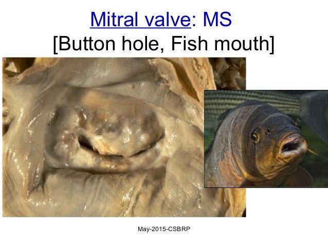
2D echo shows- calcified mitral valves
Fish mouth appearance
DIAGNOSIS: MITRAL STENOSIS WITH HEART FAILURE
Abnormal LFT and RFT are due to hypo perfusion to liver and kidney.
MITRAL STENOSIS:
Mitral stenosis is always rhuematic in origin although in older people is can be caused by heavy calcification of the mitral valve. There is also a rare form of congenital mitral stenosis.
Patient is usually asymptomatic until the orifice is < 2 cm sq. As stenosis progresses there is dilatation of left ventricle and raised left atrial pressure, leading to pulmonary congestion and breathlessness.
Atrial fibrillation is very common due to dilatation of LV
Pulmonary hypertension occurs to prevent pulmonary edema. This leads to :
Right ventricular hypertrophy and dilatation
Tricuspid regurgitation
Right heart failure
TREATMENT:
Anticoagulation to reduce the risk of embolism
B blockers
Calcium antagonists in AF
Diuretics to control pulmonary congestion
Antibiotics against infective endocardiditis.
Surgical: Mitral Balloon Valvuloplasty and Valve replacement.
ref: Davidson's principles and practice of medicine
CASE 3:
CASE 3:
https://saikiranpatnam.blogspot.com/2020/05/medicine-case.html?m=1
A 30 yr old male patient came to the OPD with chief
Bilateral pedal edema extending upto knee since 15 days
Dyspnoea on exertion since 15 days
Dry cough without expectoration since 15 day
Decreased urine output since 2 days
H/O palpitations since 1 year- persistant, ponding type
No similar complaints in past and no significant family history
GENERAL EXAMINATION
no signs of pallor, icterus, cyanosis , clubbing, lymphadenopathy.
Edema- bilateral pitting type edema extending upto knee
He belongs to TANNER STAGE 4
Vitals:
Temp- afebrile
Pulse- 124 beats/ min
RR- 20 cycles/min
BP-120/70 mmhg
SYSTEMIC EXAMINATION:
RS: Normal
GI: NO tenderness and all quadrants moving equally with respiration.
CNS:
HMF- Normal
High stepping gait-occurs due to
Decreased to power of lower limb proximal muscles-occurs due to
Two point discrimination impaired on lower limbs- occurs due to damage to
CVS:
Visible pulsation over apex and mitral areas.
Apex beat over 5th intercoastal space
Right ventricular heave is present
Jvp raised with prominent 'a' wave
S1 S2 heard with prominent p2
These findings are consistent with PULMONARY HYPERTENSION
INVESTIGATIONS:
2D echo- shows Dilated right atrium and right ventricle
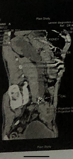
Scrotal USG- b/l scrotal sac empty
both testes not found in b/l inguinal region
PROVISIONAL DIAGNOSIS:

ACTIVE LEARNING:
A 30 yr old male patient came to the OPD with chief
Bilateral pedal edema extending upto knee since 15 days
Dyspnoea on exertion since 15 days
Dry cough without expectoration since 15 day
Decreased urine output since 2 days
H/O palpitations since 1 year- persistant, ponding type
No similar complaints in past and no significant family history
GENERAL EXAMINATION
no signs of pallor, icterus, cyanosis , clubbing, lymphadenopathy.
Edema- bilateral pitting type edema extending upto knee
- He has undescended testis ( cryptorchidism )
- Very less facial hair
- Very less pubic hair
- No axillary hair
He belongs to TANNER STAGE 4
Vitals:
Temp- afebrile
Pulse- 124 beats/ min
RR- 20 cycles/min
BP-120/70 mmhg
SYSTEMIC EXAMINATION:
RS: Normal
GI: NO tenderness and all quadrants moving equally with respiration.
CNS:
HMF- Normal
High stepping gait-occurs due to
- peroneal muscle atrophy
- peroneal nerve injury
- Spinal problem such as spinal stenosis or herniated disc
Decreased to power of lower limb proximal muscles-occurs due to
- drugs, alcohol
- thyroid disease
- osteomalacia
- inflammatory / inherited myopathies
- malignancy
- infections
- sarcoidosis
Two point discrimination impaired on lower limbs- occurs due to damage to
- posterior column-medial lemniscus pathway
- peripheral nerve
These findings point towards a possible neuromuscular disorder or myopathy which can be diagnosed by neurophysiological studies, muscle imaging and muscle biopsy
CVS:
Visible pulsation over apex and mitral areas.
Apex beat over 5th intercoastal space
Right ventricular heave is present
Jvp raised with prominent 'a' wave
S1 S2 heard with prominent p2
These findings are consistent with PULMONARY HYPERTENSION
INVESTIGATIONS:
2D echo- shows Dilated right atrium and right ventricle
Scrotal USG- b/l scrotal sac empty
both testes not found in b/l inguinal region
PROVISIONAL DIAGNOSIS:
- Pulmonary artery hypertension with right heart failure
- Hypogonadism ( tanner stage 4)
- Lower limb proximal myopathy
- oral candidiasis
- impaired glucose tolerance
Treatment given
Tab. Pantop 40 mg po/ OD
Inj. Lasix 20 mg iv/ BD
Inj. Thiamine 1amp. in 100 ml NS
Inj. Optineurin 1amp in 100 ml NS
Tab sildenafil 10mg po OD
Tab benformet plus od
On diuretic therapy and vaso dilator therapy patient got better and discharged in stable condition
Advice at discharge
Fluid (1.5 to 2 L/day) and salt (2 gm /day) restriction
Tab sildenafil 10 mg po/bd
Tab benformet po/OD for 2 weeks
Chlorhexidine oral gargles for two weeks
Oral candid paint
Diet according to Harvard plate
Work up for FSH,LH,GNRH
PULMONARY HYPERTENSION
Pulmonary hypertension is defined as mean pulmonary arterial pressure of atleast 25 mmhg at rest.
Classification of pulmonary hypertension:
- pulmonary arterial hypertension
- pulmonary venous hypertension
- pulmonary hypertension associated with diseases of respiratory system and/or hypoxemia
- pulmonary hypertension caused by chronic thromboembolic disease
- miscellaneous
Ref:F2.medium.gif
Clinical features: breathlessness, chest pain, fatigue, palpitation and syncope
Important signs include :
Investigations:
- X-ray chest
- ECG
- Doppler assessment of tricuspid regurgitant jet by transthoracic echocardiography
- Right heart catheterisation
- Signs of interstitial lung disease or cardiac, liver or connective tissue damage may suggest underlying cause.
TREATMENT:
- diuretics therapy for patients with right heart failure
- anticoagulation should be considered if there is a risk of bleeding
- digoxin may be used in patients who develop atrial tachyarrithmias
- pnuemococcal influenza vaccine should be recommended
- phosphodiesterase V inhibitors such as sildanafil may be used to enhance NO mediated vasodilatation.
Ref: Davidsons Principles and Practice of Medicine
ACTIVE LEARNING:
Case 1:
[26/05/20, 4:49:01 PM] Trishaala Reddy: Sir if we were suspecting viral myocarditis in the first case why wasn’t serology done?
[26/05/20, 4:50:23 PM] Trishaala Reddy: Other than the history of fever which the patient had a month ago what else can point towards a viral etiology
[26/05/20, 4:50:39 PM] Dr Rakesh Biswas Sir: What are the available serologies in India for viral myocarditis?
[26/05/20, 4:51:01 PM] Trishaala Reddy: Can I read and get back to you sir?
[26/05/20, 4:51:27 PM] Dr Rakesh Biswas Sir: Absolutely. There is no other way 👍
[26/05/20, 5:58:25 PM] Trishaala Reddy: Sir most of the articles say the diagnosis I mainly by clinical presentation and imaging
[26/05/20, 6:01:29 PM] Trishaala Reddy: But we do have serological tests for EBV adeno enterovirus coxsackie virus etc
[26/05/20, 6:46:42 PM] Dr Rakesh Biswas Sir: What is the sensitivity specificity of those tests?
What is the likelihood if your test comes positive for EBV then you actually have EBV (specificity aka unlikelihood of false positivity).
Similarly if you have clinical strong suspicion of EBV but it comes negative then what is the sensitivity of the test (aka false negativity)?
[26/05/20, 7:59:58 PM] Trishaala Reddy: Sir sensitivity around 85% and specificity around 100%
[26/05/20, 8:48:56 PM] Dr Rakesh Biswas Sir: Support every statement with quotes from reference links
[26/05/20, 9:02:13 PM] Trishaala Reddy: https://emedicine.medscape.com/article/222040-workup
[26/05/20, 9:12:01 PM] Dr Rakesh Biswas Sir: They haven't used cross references. Medscape is not a scientific site but a text book based drug company backed platform that is best avoided to learn anything new
[26/05/20, 9:22:23 PM] Trishaala Reddy: . “Between 85% and 90% of adolescents and adults are positive during the course of EBV infection: about 50% in the first week, and 60%-90% in the second and third[29]. However, only 50% of children aged 2-5 years are positive at any time during the course of infection, and only 10%-30% of those aged less than 2 years[2]. The rate of false negative results can therefore be high in young children, whereas false positive results are observed in only 2%-3% of the patients with autoimmune diseases[2].”
https://www.ncbi.nlm.nih.gov/pmc/articles/PMC3782265/
[26/05/20, 10:04:04 PM] Dr Rakesh Biswas Sir: 👍
Are there any studies to suggest that EBV could have caused his myocarditis?
[26/05/20, 10:12:08 PM] Trishaala Reddy: Sir there is an article which says it is a rare cause but around 15 cases were reported
[26/05/20, 10:13:24 PM] Trishaala Reddy: https://www.ncbi.nlm.nih.gov/m/pubmed/20561866
[26/05/20, 10:19:57 PM] Trishaala Reddy: There are also 2 other cases but the age of presentation is much younger
https://www.ncbi.nlm.nih.gov/m/pubmed/10941492
https://www.ncbi.nlm.nih.gov/pmc/articles/PMC6907365/
[27/05/20, 9:26:54 PM] Dr Rakesh Biswas Sir: Nice find 👍👏👏
[27/05/20, 10:18:12 PM] Dr Rakesh Biswas Sir: Share your learning insights from the article
[27/05/20, 10:24:45 PM] Trishaala Reddy: Sir I have learned that most often dilated cardiomyopathy occurs due to idiopathic reasons.
We usually rule out infarction and hypertension as a cause.
Viral myocarditis leading to dilated cardiomyopathy is well known cause. Most commonly it is due to adenovirus.
EBV is a very rare cause of myocarditis.
Mechanism of damage to the myocardium by the virus maybe direct , toxic or by autoimmune mechanism.
Treatment is the standard heart failure therapy.
[27/05/20, 10:26:33 PM] Trishaala Reddy: Accurate diagnosis can be made by endomyocardial biopsy and PCR for detection is viral genome.
[27/05/20, 10:27:55 PM] Trishaala Reddy: Sir so is our patient is it not very necessary to know the virus causing myocarditis as the treatment is usually standard ?
[27/05/20, 10:31:16 PM] Dr Rakesh Biswas Sir: Yes nothing can be done currently for whatever virus it may have been.
Currently it is only the treatment of heart failure for him.
Tell me what is the efficacy of neprelysin inhibitors and the combination with ARB that he is receiving?
Also ask the treating PG and interns
[28/05/20, 10:48:01 AM] Trishaala Reddy: Good morning sir.
Sir sacubitril is a neprilysin inhibitor and valsartan Is an ARB.
Valsartan decreases aldosterone, increases natriuresis and causes vasodilation and reduces BP. Sacubitril inhibits neprilysin (neprilysindeactivates natriuretic peptides). This leads to increased levels of BNP and bradykinin which cause myocardial relaxation and reduced hypertrophy.
Hence sacubitril/valsartan can be used as a combination drug in heart failure patients
[28/05/20, 10:48:53 AM] Trishaala Reddy: Ref: https://www.ncbi.nlm.nih.gov/pmc/articles/PMC6426571/
[28/05/20, 4:21:42 PM] Dr Rakesh Biswas Sir: Yes these are the mechanisms of action. What is the efficacy of ARNI over placebo?
[28/05/20, 9:35:00 PM] Trishaala Reddy: “Efficacy and Safety of LCZ696, a First-in-Class Angiotensin Receptor Neprilysin Inhibitor, in Asian Patients With Hypertension: A Randomized, Double-Blind, Placebo-Controlled Study”
Ref: https://www.researchgate.net/publication/259826466_Efficacy_and_Safety_of_LCZ696_a_First-in-Class_Angiotensin_Receptor_Neprilysin_Inhibitor_in_Asian_Patients_With_Hypertension_A_Randomized_Double-Blind_Placebo-Controlled_Study
•PATIENT/ PROBLEM/POPULATION:
Asian patients aged =/> 18 yrs with hypertension
•INTERVENTION:
LCZ 696 ( sucabitril valsartan sodium hydrate)
100 mg ( n= 100)
200 mg ( n = 101)
400 mg ( n= 96)
•COMPARISON:
Placebo (n=92)
•OUTCOME:
Reduction in systolic BP, diastolic BP and pulse pressure were significantly greater with all doses of LCZ 696 than with placebo.
It was well tolerated and no cases of angioedema were reported.
In conclusion LCZ 696 is an effective treatment for hypertension in Asian population and is safe and well tolerated.
[28/05/20, 9:38:28 PM] Dr Rakesh Biswas Sir: Very well done. This was more of phase 2 trial.
Now check out ARNI vs ACEI or ARB alone
[29/05/20, 12:26:51 AM] Trishaala Reddy: “The Prospective Comparison of ARNI with ACEI to Determine Impact on Global Mortality and Morbidity in Heart Failure (PARADIGM-HF) trial “
Ref: https://www.ncbi.nlm.nih.gov/pmc/articles/PMC6426571/
•PATIENT/ PROBLEM/POPULATION:
8399 patients with HFrEF NYHA II-IV
•INTERVENTION:
Sacubitril/Valsartan ( ARNI )
•COMPARISON:
Enalapril (ACEi)
•OUTCOME:
Deaths due to cardiovascular causes were 13.3% in the sacubitril/valsartan group compared to 16.5% in the enalapril group.
Hospitalizations for HF were 12.8% and 15.6% of patients receiving sacubitril/valsartan and enalapril respectively.
In addition, the benefit with respect to cardiovascular mortality was consistent irrespective of age, sex, ethnicity, ejection fraction and NYHA class, with no impact on the incidence of non-cardiovascular death, meaning that the mortality benefit was due to decreased cardiovascular risk. Further analysis revealed that the hazard for both sudden death and death due to worsening HF was significantly reduced by treatment with sacubitril/valsartan
Case 2:
[30/05/20, 5:10:19 PM] Trishaala Reddy: Maam the reason for abnormal RFT and LFT is hypo perfusion to the kidney and liver or is there any other mechanism ?
[30/05/20, 5:12:18 PM] Trishaala Reddy: Also ma’am since rheumatic is the most common cause of mitral stenosis...how did we rule it out in this case?
[30/05/20, 5:14:10 PM] Radha Maam Kims: Can you send the reports?
[30/05/20, 5:15:17 PM] Radha Maam Kims: Yeah but the patient came in a late stage ma... so basically even if we do a biopsy... all we can find is fibrotic changes. Aschoff bodies are seen in acute rheumatic fever only
[30/05/20, 5:15:57 PM] Radha Maam Kims: Based on the mitral valve morphology in 2d echo... we thought it could most probably be secondary to ms
[30/05/20, 5:16:35 PM] Radha Maam Kims: We had so many heart failure cases recently so I don’t want to get confused here... as far as I remember it is due to hypoperfusion
[30/05/20, 5:23:45 PM] Trishaala Reddy: Maam don’t we chech for ASO titre?
[30/05/20, 5:24:37 PM] Trishaala Reddy: https://himabindu5.blogspot.com/2020/05/hello-everyone.html
[30/05/20, 5:32:44 PM] Radha Maam Kims: Aso titer has very low sensitivity and specificity
[30/05/20, 5:33:18 PM] Radha Maam Kims: And at this point where the patient went into cardiogenic shock... knowing the diagnosis will not change the treatment course
[30/05/20, 5:33:42 PM] Trishaala Reddy: Ohhkk thank you Maam

Comments
Post a Comment